Fractures
Objectives
Upon finishing this module, the student will be able to:
- Describe pediatric bony anatomy.
- Identify a Torus or Buckle fracture and describe its clinical significance.
- List the different classifications of Salter-Harris Fractures I-V.
- Describe the clinical significance and treatment options for physeal fractures.
Contributors
Update Authors: Karina Hofstee, MD, MS; and Nidhi "Nina" Singh, MD.
Original Author: Todd Peterson, MD.
Update Editor: Lisa Armstrong, MD.
Last Updated: July 2024
Introduction
Orthopedic trauma accounts for 10-15% of emergency department (ED) visits in pediatric hospitals.1 Injury frequency increases as children become more mobile and active. The immaturity of the pediatric skeletal system often causes significant differences from adults in terms of fracture patterns and imaging findings. Subtle radiographic findings can be associated with fractures and misinterpretation of a radiograph can lead to lifelong functional deficits, including limb-length discrepancies. It is important to maintain a high index of suspicion for a fracture in any child who has a history of a traumatic injury, presenting with focal bony pain or refusal to use a limb.
Pediatric Bony Anatomy
Long bones in children are composed of the diaphysis (shaft), metaphysis (flared area), physis (growth plate), and epiphysis (secondary ossification center). Pediatric bones have a lower tensile strength related to attached ligaments than skeletally mature bone, this results in higher rates of fractures from mechanisms that might otherwise cause a sprain or dislocation in adults.1 There are two main anatomic considerations for the pediatric bone:
- First, the periosteum of the pediatric bone is more metabolically active, thicker, and stronger than adult periosteum.2 This promotes callus formation and bone remodeling during healing, limiting fracture displacement, which results in unique fracture patterns not seen in adults, such as torus, buckle, and greenstick fractures.
- The second anatomic consideration is the presence of the growth plate, or physis. The physis is a hyaline cartilage plate where bone growth primarily occurs.

Figure: Basic anatomy of pediatric long bones. Image courtesy of Aneta Kecler-Pietrzyk. Radiopaedia.org. Used under the creative commons license.
The position of a fracture relative to the metaphysis, physis, and epiphysis has a tremendous impact on the clinical significance and prognosis of the fracture. Growth plate injuries are often characterized by the Salter-Harris classification system, discussed below.
As with any presentation of acute traumatic injury, initial management should be focused on the “ABCs”, first assessing the patient’s airway, breathing, and circulation. Next, management should include treatment of pain and evaluation of the injury, with the goal of preserving long-term function. Particular attention should be paid to circulation as it relates to orthopedic injuries. Check neurovascular status of the affected limb by assessing motor and sensory function as well as pulses and capillary refill distal to the suspected fracture site. Other signs of complicated orthopedic injuries include open fractures that lead to an increased risk of infection, and fractures that involve the growth plate.1 The physical exam of the affected extremity should include inspection, palpation, range of motion, neurovascular function, and examination of the proximal and distal joints.
It is important to consider that the ABCs always come first in the evaluation of the trauma patient. A common mistake is to focus prematurely on a deformed extremity and miss another life-threatening injury. A quote to remember is "life over limb."
Plain film radiographs are typically sufficient to make a diagnosis of fracture. Localization of the injury may be challenging clinically, so it is common for the joint above and below the suspected area of injury to be imaged as well. The Image Gently Campaign recommends using Pediatric protocols to decrease radiation exposure. Computed Tomography (CT) is often not indicated for diagnosis of most pediatric fractures in the ED due to the risk of radiation exposure, but may be used for evaluation of complex joint or spinal injuries.
Torus or Buckle Fractures
This type of fracture is characterized radiographically by a small bulging of the cortex. It most commonly occurs after an axial load or compression injury in young children. The most common location is at the junction of the dense bone of the diaphysis and the more porous, immature bone of the metaphysis.2 Treatment of buckle fractures consists of splinting in the emergency room (ER) with outpatient orthopedic follow-up. Fracture reduction is unnecessary, and immobilization is typically only needed for less than one month.
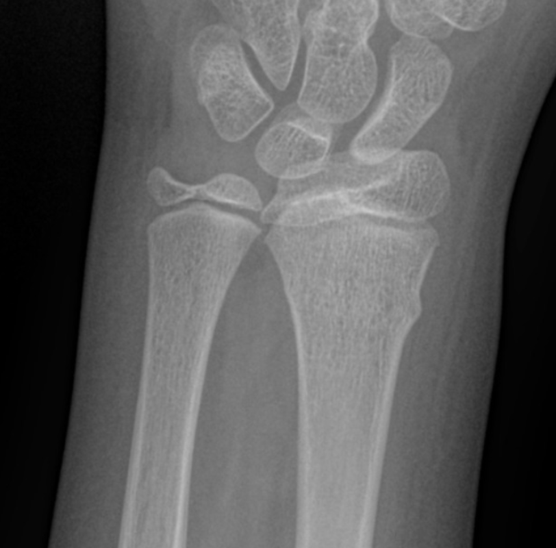
Figure: Buckle fracture. Image courtesy of Mostafa Elfeky. Radiopaedia.org. Used under the creative commons license.
Greenstick Fractures
Due to the thick and active periosteum in pediatric bones, the bone often starts to bend before it breaks. Greenstick fractures show a pattern of bowing where the periosteum remains intact on the concave side of the bow and is torn on the convex side, like breaking a small, healthy, green branch.5 When a large amount of angulation is present, they may need to be reduced in the ED. During reduction, the fracture may need to be exaggerated and completed to achieve anatomic alignment and avoid loss of function.1 Once aligned, the fracture should be immobilized and orthopedic follow-up provided.

Figure: Greenstick fracture. Image courtesy of Leonardo Lustosa. Radiopaedia.org. Used under the creative commons license.
Toddler's Fractures
Toddler's fractures occur in children aged nine-36 months. This is usually an oblique or spiral, non-displaced fracture of the distal tibia that occurs after a minor fall, though sometimes there is no reported history of trauma.1 The physical exam may be normal outside of a refusal to bear weight on the affected extremity, and/or tenderness with manipulation. Radiographs may appear normal initially, with signs of bone healing at the fracture line only visible seven-14 days after the injury. Therefore, even in cases with an initial "normal" x-ray, if the child is refusing to bear weight the extremity should be immobilized and orthopedic follow-up provided.

Figure: Toddler's fracture. Image courtesy of Jeremy Jones. Radiopaedia.org. Used under the creative commons license.
Physeal (Growth Plate) Fractures
The Salter-Harris classification describes physis fractures with grades I-V involving the growth plate. Most growth plate injuries occur in the upper limbs, particularly during periods of rapid growth such as adolescence.1 The distinctions between the types are significant as it impacts treatment and prognosis of the injury. The SALTR mnemonic is useful for remembering the different classifications: straight across, above, lower or below, through, crush.

Figure: Salter-Harris classification of growth plate injuries. Image courtesy of Matt Skalski. Radiopaedia.org. Used under the creative commons license.
Salter-Harris Type 1
A Salter-Harris type 1 fracture separates the metaphysis from the epiphysis and causes a widening of the physeal space. It represents 6% of Salter-Harris fractures.9 It is often diagnosed clinically based on point tenderness at the physis on exam. There may be no radiographic abnormality on the initial x-ray. The best management plan is to immobilize the joint with a splint and have the patient follow up with an orthopedist in one week for repeat imaging. Long-term prognosis is good and surgery is typically not indicated. If there is displacement of the epiphysis, urgent reduction will be needed.

Figure: Salter-Harris I fracture. Image courtesy of Mauricio Macagnan. Radiopaedia.org. Used under the creative commons license.
Salter-Harris Type II
A Salter-Harris type II fracture involves the physis and the metaphysis. This is the most common Salter-Harris fracture, accounting for 75% of all growth plate injuries.9 This fracture type usually carries a good prognosis, rarely resulting in any functional deformity. Fractures with angulation and displacement may need reduction. Acceptable amounts of angulation will vary depending on the age of the patient and location of the fracture.

Figure: Salter-Harris II fracture. Image courtesy of Leonardo Lustosa. Radiopaedia.org. Used under the creative commons license.
Salter-Harris Type III
A Salter-Harris type III fracture involves the physis and the epiphysis, making it an intra-articular fracture. This accounts for 7-10% of growth plate injuries and typically occurs after the age of ten due to partial fusion of the physis.9 The prognosis is often poorer as the proliferative and reserve zones of the growth plate are interrupted. Early reduction into anatomic alignment is crucial for long-term outcomes, and orthopedics should be consulted in the ER for all displaced fractures. Surgical treatment may be needed to achieve proper reduction and preservation of growth plate integrity as growth plate injury can lead to limb-length discrepancies. Non-displaced fractures should be splinted and with expeditious orthopedics follow-up.

Figure: Salter-Harris III fracture. Image courtesy of Pediatric Imaging. Pediatricimaging.org. Used under the creative commons license.
Salter-Harris Type IV
A Salter-Harris type IV fracture involves the articular surface of the epiphysis, going across the physis and through the metaphasis, making it an intra-articular fracture. This accounts for roughly 10% of all growth plate injuries and typically carries a poor prognosis.9 Early reduction is crucial and orthopedics should be consulted in the ER for all displaced fractures, as surgical treatment is often necessary.

Figure: Salter-Harris IV fracture. Image courtesy of Hisham Alwakkaa. Radiopaedia.org. Used under the creative commons license.
Salter-Harris Type V
A Salter-Harris type V fracture involves an axial loading mechanism with compression of the physis and disruption of the germinal matrix.2 This is relatively rare and accounts for less than 1% of growth plate injuries. Proper diagnosis is dependent upon recognition of a mechanism of a significant axial load force on the extremity. Radiographic changes are subtle and involve narrowing of the physis with a joint effusion. Misdiagnosis is common and prognosis is poor, as the compressive force may cause premature closure of the physis and result in growth arrest of the bone. Typically no significant displacement is present, but orthopedics should be consulted to arrange follow-up in anticipation of growth disruption.
The majority of pediatric fracture patients will be immobilized in a splint and discharged home with timely orthopedic follow-up. Emergent orthopedic consultation is required for fractures that are open, significantly displaced, or have neurovascular compromise. Urgent orthopedics evaluation may be needed for fractures involving the growth plate.
It is important to provide adequate analgesia both in the ER and at home. Immobilization helps provide pain control and prevent further injury. Ibuprofen and acetaminophen are first-line for pain control at home and adequate for most cases.
- Maintain a high index of suspicion for fracture in any child with acute functional limitation in an extremity.
- Imaging the joint above and below the suspected injury is crucial to ensure no fractures are missed.
- "Normal" imaging does not exclude a fracture (i.e. Type I supracondylar, toddler's fracture, Salter-Harris type I fracture).
- It is acceptable to splint a patient and have them follow-up for repeat imaging if the child has persistent pain or a functional limitation.
- Remodeling and fracture union are robust in the pediatric skeleton, thus less operative repair is needed than in adult fractures.
- Early closure or disruption of the growth plate can lead to permanent deformity and limb length discrepancies.
- Always document neurovascular status before and after splint placement.
After your thorough history and physical exam including neurovascular status, you obtain radiographs of your patient's injured extremity. In addition to the wrist and forearm, you also obtain radiographs of the elbow on the affected side, which were normal.

Figure: AP and lateral radiographs. Image courtesy of Pediatric Imaging. Pediatricimaging.org. Used under the creative commons license.
The radiographs show distal radius and ulna buckle or torus fractures. As these are stable fractures that do not involve the growth plate, you apply a splint to immobilize the arm and provide a referral to the orthopedic clinic next week. The patient feels better after splint placement and receiving a dose of ibuprofen.
- Thompson R, Hannon M, Lee L. Musculoskeletal Trauma. Fleisher & Ludwig's Textbook of Pediatric Emergency Medicine, 8th ed. Wolters Kluwer. 2021.
- Mathison D, Agrawal D. General Principles of Fracture Management: Fracture Patterns and Description in Children. UpToDate. 2023.
- Kecler-Pietrzyk A. Normal Femur, Tibia, and Fibula X-Rays (1-Year-Old). Radiopaedia.org. 2017.
- El-Feky M. Buckle Fracture - Distal Radius. Radiopaedia.org. 2022.
- Rang M, Pring M, Wenger D. Rang's Children's Fractures, 3rd ed. Lippincott Williams & Wilkins. 2005.
- Lustosa L. Radial Greenstick Fracture. Radiopaedia.org. 2023.
- Jones J. Toddler Fracture. Radiopaedia.org.
- Skalski M. Salter-Harris Illustrations. Radiopaedia.org. 2014.
- Qureshi P, Gaillard F. Salter-Harris Classification. Radiopaedia.org. 2008.
- Macagnan M. Salter-Harris Type I. Radiopaedia.org.
- Lustosa L. Salter-Harris Type II Fracture of Distal Tibia. Radiopaedia.org. 2022.
- Salter-Harris Fracture. Pediatric Radiology Reference. Pediatric Imaging.
- Glick Y, Alwakkaa H. Salter-Harris Type IV Fracture. Radiopaedia.org. 2017.
- Torus Fracture. Pediatric Radiology Reference. Pediatric Imaging.